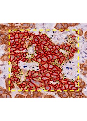
The Aperio Membrane Algorithm detects membrane staining for individual tumor cells in a selected region, and quantifies the intensity and completeness of the membrane staining. Use the Aperio Membrane Algorithm to measure expression of IHC biomarkers specifically within the cell membrane, while excluding staining in other subcellular compartments (cytoplasm, nucleus).
Learn more
Highly specific analysis
Tunable algorithm settings let you precisely define parameters for cells to be analyzed, including size, shape and membrane staining intensity and completeness. Analyze only the cells that are of interest for your research.
Analyze the right tissue
Analysis can be run on fields of view, annotated regions, or whole slides. Pre-process slides with Aperio GENIE for auto-selection of tumor, removing the need for manual annotation.
Scoring that fits your preferences
You set the parameters that define scoring classes (0, 1+, 2+, 3+) so your algorithm can be adjusted for variations in staining due to reagent choice or slide preparation.
Easy to review results
Analyzed regions are overlaid with a color coded mark-up in Aperio ImageScope for simple visual review of the data. Aperio Membrane Algorithm assigns an overall score, but also shows the absolute percentage of cells in each scoring class, so you can make a determination in borderline cases.


